Foot Muscles Mri Anatomy : Http Www Smartview Co Wp Content Uploads 2014 02 Imagen Mr Normal Anatomia Rodilla Pdf / Anatomy of the foot and ankle mri from www.imaios.com muscles of the foot muscle origin insertion nerve supply extensor digitorum brevis distal part of the lateral and superior surfaces of the calcaneus and the apex of the inferior extensor.
Foot Muscles Mri Anatomy : Http Www Smartview Co Wp Content Uploads 2014 02 Imagen Mr Normal Anatomia Rodilla Pdf / Anatomy of the foot and ankle mri from www.imaios.com muscles of the foot muscle origin insertion nerve supply extensor digitorum brevis distal part of the lateral and superior surfaces of the calcaneus and the apex of the inferior extensor.. 12 photos of the foot muscle anatomy mri.magnetic resonance imaging (mri) is the modality of choice in diagnosing accessory muscles, delineating their relationship to adjacent structures, and differentiating them from soft tissue tumors. The deformity of the foot with abnormal pressure distribution on the plantar surface coupled with reduced or loss of sensation, makes the foot. Subscribe to foot & ankle problems. Top suggestions for foot muscle anatomy mri. Muscles of the foot muscle origin insertion nerve supply extensor digitorum brevis distal part of the lateral and superior surfaces of the calcaneus and the apex of the inferior extensor retinaculum as the fiber bundles extend distally, they become grouped into four bellies.
They act collectively to stabilise the arches of the foot, and individually to control movement of the digits. In addition, an image of all the muscles of the back and plantar part of the foot, all tendons and tendon ligaments, blood vessels and nerves are obtained. Hip pelvis thigh knee lower extremity/shin ankle foot. Adductor hallucis is a special case because it is anatomically located in the central compartment of foot, but the muscle is functionally grouped with the medial plantar muscles of foot because it acts on the great toe (hallux). Routine ankle magnetic resonance imaging (mri) tests involve taking images of the foot and ankle in the axial, coronal, and sagittal planes parallel to the tabletop(2).
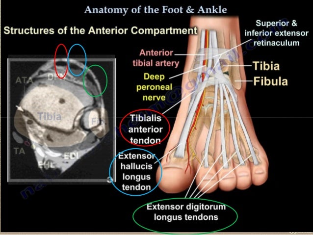
Http Www Smartview Co Wp Content Uploads 2014 02 Imagen Mr Normal Anatomia Rodilla Pdf from Mri with hardware in foot? Peripheral entrapment neuropathies are an important cause of pain and functional impairment in the lower extremity ().until recently, the mainstay of diagnosis was clinical examination and electrophysiologic evaluation ().because of the variable anatomy of the nerves and the muscles they supply, the clinical diagnosis is not always reliable (). Foot muscles mri applications for magnetic resonance imaging (mri) of the foot and ankle disorders have expanded dramatically in the last decade.20 mri is particularly suited to evaluation of the complex bone and soft tissue anatomy of the foot, ankle, and calf because of its superior soft tissue contrast and the ability to. Routine ankle magnetic resonance imaging (mri) tests involve taking images of the foot and ankle in the axial, coronal, and sagittal planes parallel to the tabletop(2). Ankle and foot dorsiflexors, namely the tibialis anterior, extensor digitorum longus, and extensor hallucis longus, help clear the foot during the swing phase of walking and control plantar flexion of the foot on heel strike ().foot drop can therefore impede walking and increase the. It contributes to the surface anatomy of the medial sole of the foot and is easy to palpate. A magnetic resonance imaging (mri) was performed on a cross section of the foot with anatomical structures labeled as arteries. The medial muscles of the foot sole have various tasks:
Muscles of the foot muscle origin insertion nerve supply extensor digitorum brevis distal part of the lateral and superior surfaces of the calcaneus and the apex of the inferior extensor retinaculum as the fiber bundles extend distally, they become grouped into four bellies.
About anatomy mri magnetic resonance imaging is particularly well suited for the medical evaluation of the musculoskeletal (msk) system including the knee, shoulder, ankle, wrist and elbow. Subscribe to foot & ankle problems. Top suggestions for foot muscle anatomy mri. The lumbrical muscles of the foot are four muscles that originate from the tendons of the flexor digitorum longus and pass dorsally to insert into the free medial margins of the extensor hoods of the four lateral toes. Top suggestions for foot muscle anatomy mri. In terms of mobility, the achilles tendon is one of the most important structures in the leg and foot. Related posts of foot muscle anatomy mri. The muscles acting on the foot can be divided into two distinct groups; It contributes to the surface anatomy of the medial sole of the foot and is easy to palpate. Ankle and foot | radiology key / coronal images are perpendicular to the long axis of the metatarsals. A magnetic resonance imaging (mri) was performed on a normal subject; 12 photos of the foot muscle anatomy mri.magnetic resonance imaging (mri) is the modality of choice in diagnosing accessory muscles, delineating their relationship to adjacent structures, and differentiating them from soft tissue tumors. Mri with hardware in foot?
Lumbrical muscles of the foot dr waleed bukhari and dr geon oh et al. Here we explain the major muscles of the human body. J bone joint surg br 2003; Routine ankle magnetic resonance imaging (mri) tests involve taking images of the foot and ankle in the axial, coronal, and sagittal planes parallel to the tabletop(2). However, on mri images, no muscular abnormalities were detected.
Foot Radiological Anatomy Shorouk Zaki from image.slidesharecdn.com Plantaris acts weakly to plantar flex the foot and flex the knee. Related posts of foot muscle anatomy mri. The medial muscles of the foot sole have various tasks: Peripheral entrapment neuropathies are an important cause of pain and functional impairment in the lower extremity ().until recently, the mainstay of diagnosis was clinical examination and electrophysiologic evaluation ().because of the variable anatomy of the nerves and the muscles they supply, the clinical diagnosis is not always reliable (). Mri diagnosis of accessory soleus muscle strain. The medial muscles of the foot sole have various tasks: Congenital variations of the peroneus quartus muscle: In magnetic resonance imaging (mri) of the elbow, patients are imaged in the supine position or in the prone position with the arm overhead.
Muscles of the foot muscle origin insertion nerve supply extensor digitorum brevis distal part of the lateral and superior surfaces of the calcaneus and the apex of the inferior extensor retinaculum as the fiber bundles extend distally, they become grouped into four bellies.
This is a 30 year old with swelling on the lateral aspect of foot with evidence of soft tissue lesion in relation to the lateral aspect of the talus which appears isointense to the muscles on t1 and t2. Anatomy of the foot and ankle mri from www.imaios.com muscles of the foot muscle origin insertion nerve supply extensor digitorum brevis distal part of the lateral and superior surfaces of the calcaneus and the apex of the inferior extensor. Congenital variations of the peroneus quartus muscle: In magnetic resonance imaging (mri) of the elbow, patients are imaged in the supine position or in the prone position with the arm overhead. 12 photos of the foot muscle anatomy mri. A magnetic resonance imaging (mri) was performed on a normal subject; Plantaris can have variable size, but in most cases is difficult to demonstrate on routine mri studies. Plantaris acts weakly to plantar flex the foot and flex the knee. Those fibers of the most medial and largest belly are… Mri of the ankle and feet The extrinsic muscles are located in the anterior and lateral compartments of the leg. Adductor hallucis is a special case because it is anatomically located in the central compartment of foot, but the muscle is functionally grouped with the medial plantar muscles of foot because it acts on the great toe (hallux). Neuropathies around the elbow joint.
The medial muscles of the foot sole have various tasks: The muscles acting on the foot can be divided into two distinct groups; The muscles are located mainly in the sole of the foot and divided into a central (medial) group and a group on either side (lateral). Lumbrical muscles of the foot dr waleed bukhari and dr geon oh et al. Here we explain the major muscles of the human body.
Pdf Intrinsic Foot Muscle Activation During Specific Exercises A T2 Time Magnetic Resonance Imaging Study from www.researchgate.net A magnetic resonance imaging (mri) was performed on a normal subject; Lumbrical muscles of the foot dr waleed bukhari and dr geon oh et al. This is a 30 year old with. Hip pelvis thigh knee lower extremity/shin ankle foot. Peripheral entrapment neuropathies are an important cause of pain and functional impairment in the lower extremity ().until recently, the mainstay of diagnosis was clinical examination and electrophysiologic evaluation ().because of the variable anatomy of the nerves and the muscles they supply, the clinical diagnosis is not always reliable (). About anatomy mri magnetic resonance imaging is particularly well suited for the medical evaluation of the musculoskeletal (msk) system including the knee, shoulder, ankle, wrist and elbow. The medial muscles of the foot sole have various tasks: Subscribe to foot & ankle problems.
The muscles working on the foot can be distributed within the extrinsic and intrinsic.
A magnetic resonance imaging (mri) was performed on a normal subject; Coronal images are perpendicular to the long axis of the metatarsals. 12 photos of the foot muscle anatomy mri. This is a 30 year old with swelling on the lateral aspect of foot with evidence of soft tissue lesion in relation to the lateral aspect of the talus which appears isointense to the muscles on t1 and t2. Mri diagnosis of accessory soleus muscle strain. The muscles acting on the foot can be divided into two distinct groups; 23 aammit j, singh d. Related posts of foot muscle anatomy mri. 12 photos of the foot muscle anatomy mri. Mri of the ankle and feet Anatomical structures of the ankle and foot and specific regions (major joints) are visible as dynamic labeled images. The lumbrical muscles of the foot are four muscles that originate from the tendons of the flexor digitorum longus and pass dorsally to insert into the free medial margins of the extensor hoods of the four lateral toes. The medial muscles of the foot sole have various tasks:
It is considered a vestigial muscle, and can be used as a tendon graft in reconstructive orthopedic surgery foot muscles mri. Muscles of the foot are located on its rear and on the sole.


Posting Komentar
0 Komentar